One. Experimental Principles
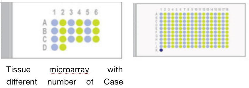
Tissue microarray technology is a new molecular biology technology based on morphology developed in recent years. It is an analysis tool with high-throughput and multi-samples. It is a high-throughput tissue slice made by arranging dozens or even thousands of micro tissue slices neatly on a slide to form a microarray. By hybridizing the nucleic acid probe or antibody probe labeled with a specific gene with it, the expression of the gene in different tissues can be deteced. Therefore, tissue microarray is the integration of traditional nucleic acid in situ hybridization or immunohistochemistry experiments. Nucleic acid in situ hybridization or immunohistochemistry can detect the expression of one gene in one tissue at a time, while tissue microarray can detect the expression of one gene in multiple tissues at a time.
Two. Application Introduction
1. Cell phenotype analysis
Using immunohistochemistry to detect various indexes of different tissue samples on the tissue chip, compared with the complete large tissue sections, the tissue chip constructed from different parts can provide a reliable high-throughput immunohistochemical phenotype analysis system.
2. Combined application with gene chip
Tissue microarray technology can also detect the expression of several or dozens of gene products at the same time, which can be used to find the regulation of various genes in various tissue samples, and then obtain valuable experimental results according to these different regulation conditions.
3. New gene targets screening
Using tissue microarray to analyze each candidate gene can find the most potential target gene for new drugs or inhibitors, or find proto oncogenes or new genes encoding signal transduction molecules.
4. Microstructural and pathological Atlas
Various micro histological and pathological maps can be prepared as needed.
5. Antibody screening
In the research of various diseases, disease-related antibodies and probes are essential research tools. The basic method of antibody and probe testing is to examine a large number of positive and negative tissues from different sources. In this regard, a large number of single sections need to be made in traditional pathological methods. However, if using tissue chip technology, it can be done with one experiment.
6. Individualized tumor treatment
Tissue microarray can be used to screen a large number of tumor tissue samples to determine the specific treatments for specific tumour.
Three. Experimental Methods
1. Select the tissues to be studied.
2. Mark the area to be studied after testing.
3. Use tissue chip device to arrange the marked tissues on the blank wax block mold according to the design.
4. Slice the arranged wax block with a slicer to get the tissue chips.
Four. Sample delivering requirements
Sample type | Sample requirements | Preservation conditions | Delivery conditions | Note |
Fresh tissue | For the fresh tissue, it should be washed the blood away with PBS and preserve in -80°C immediately | At -80°C | With dry ice | All samples need to be uniquely marked and the markings are clearly identifiable |
Regular fixed tissue | The volum of the regular tissue is within 2cm*2cm*0.5cm in general. Fixation is to keep the original shape of the tissue as much as possible and clean up the blood and excess parts; the volum of fixative fluid should be at least 10 times of the tissue, and the container should be big enough to hold the tissue without squeezing it. The type of fixative fluid and the fixed duration should be marked | At ambient temperature | At ambient temperature | |
Special fixed tissue | For tissue like testicle, eyeball, spinal cord, muscle, and so on: the correspondent special fixative fluid is recommended for fixation to ensure the fixing effect. | |||
Paraffin embedded tissue | The tissue should be embedded in a standard paraffin embedding cassette, and paraffin wax should be evenly bonded to the tissue with no crack; the thickness of the wax block is determined by the number of slices required, and the effective thickness should be at least 0.1 cm. Provide the paraffin embedded tissue made within six months, otherwise the antigens of the paraffin may be lost and fail to detect the protein when performing immunohistochemistry. | At ambient temperature | At ambient temperature | |
Tissue section | It needs to pick up the section with anti-drop slides and mark the thickness of the section, and the temperature and duration of baking. The tissue should be near the lower third of the slice | At 4°C | With ice bag | |
Cell climbing sheet | Use the special cell climbing sheet for the orifice plate. After the cell climbing sheet is fixed, wash it with PBS for 2 to 3 times, store it in PBS, and seal the orifice plate with sealing film. Each climbing sheet shall have a unique mark on the orifice plate cover and electronic grouping information to prevent the mark from being blurred during transportation. | At ambient temperature | At ambient temperature | |
Plant example | Fix the fresh tissue with FAA and its degree of lignification is not high; for thinner blades, if parallel blade slicing is required, it is difficult to ensure complete cutting; the diameter of the rhizome should be larger than 1 mm. | At ambient temperature | At ambient temperature | |
Antibody | There should be clear marks to identify the antigens on the containers and the instructions should be attached. The amount of antigens should be larger than the experiment required. For tissue sections and tissue chips, calculate the amount of antibody according to a section of 100 μl after dilution; for the round coverslips of the 24 well plate cell climbing slide, calculate the amount of antibody according to a section of 100 μl after dilution; for the round coverslips of the 12 well plate cell climbing slide, calculate the amount of antibody according to a section of 400 μl after dilution | At -20°C | With ice bag or dry ice |
Five. Case Display

Six. Common Problems
1. The slices of tissue chip should not be sliced continuously. Each slice should be frozen with ice once;
2. If some tissues are too brittle, wipe them with an atomizer or a wet towel with warm water and slice them;
3. The water temperature for picking up the slice is about the melting point of the paraffin minus 15 ℃. Generally, the water temperature is controlled between 42-45 ℃. If the water is too hot, the tissue will be scattered, and if the water too cold, the wax piece will be wrinkled;
4. The effect of slicing tissue chip with flat push slicer is better;
5. Disposable blades are preferred;
6. The slice is generally 4um.
Seven.Service Process


